

Eliminating Acoustic Feedback in High-Gain Custom Earmolds Using Low-Viscosity Addition Silicone

1. Clinical Baseline & Patient Presentation
A 48-year-old female with bilateral, severe-to-profound sensorineural hearing loss presented for the refitting of her custom Behind-The-Ear (BTE) hearing aids. The patient required a high-gain amplification prescription, particularly in the high-frequency spectrum, to improve speech discrimination scores.
Due to the high-output requirements of her prescription, the success of the rehabilitation depended entirely on fabricating a pair of custom acrylic earmolds that could establish an absolute, hermetic acoustic seal within the external auditory canal. The target depth for the impression was set past the second anatomical bend of the canal to ensure optimal retention and acoustic isolation.
2. The Audiological Challenge & Technical Bottlenecks
The primary clinical obstacle centered on the patient's highly sensitive, narrow, and slightly tortuous ear canal anatomy. Previous attempts at impression-taking using conventional materials at an outside clinic resulted in severe fit issues and patient discomfort. The technical bottlenecks identified included:
- Acoustic Feedback (Whistling): The patient's previous earmolds suffered from micro-gaps caused by the linear shrinkage of traditional condensation-cure (C-silicone) putties. Under high-gain amplification, sound leaked through these gaps, causing continuous, distressing acoustic feedback.
- Tissue Displacement and Distortion: High-viscosity, hand-kneaded impression putties often require significant manual pressure during injection. In this patient's narrow canal, this pressure dilated the soft cartilaginous tissue artificially, resulting in an oversized impression and a final earmold that caused painful pressure necrosis points within 2 hours of wear.
- Tear and Elongation Risks: The deep canal past the second bend presented distinct bony undercuts. Standard alginates or low-tensile silicones risked tearing or stretching during extraction, compromising the anatomical accuracy of the aperture rim and helix areas.
3. Clinical Justification for Advanced A-Silicone Selection
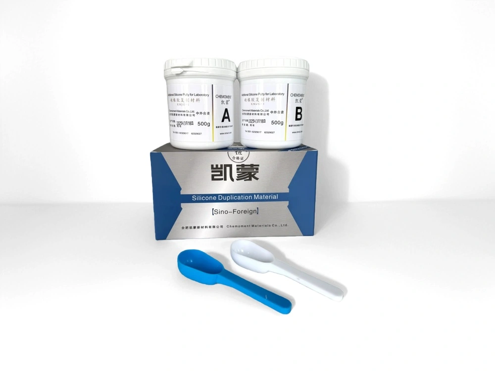
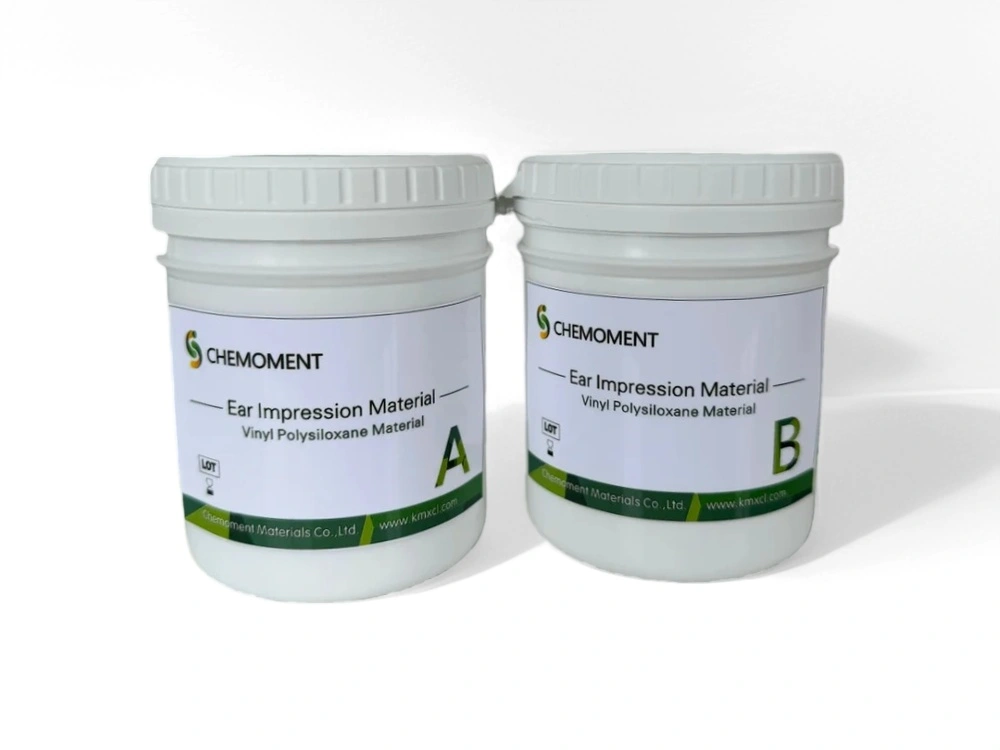
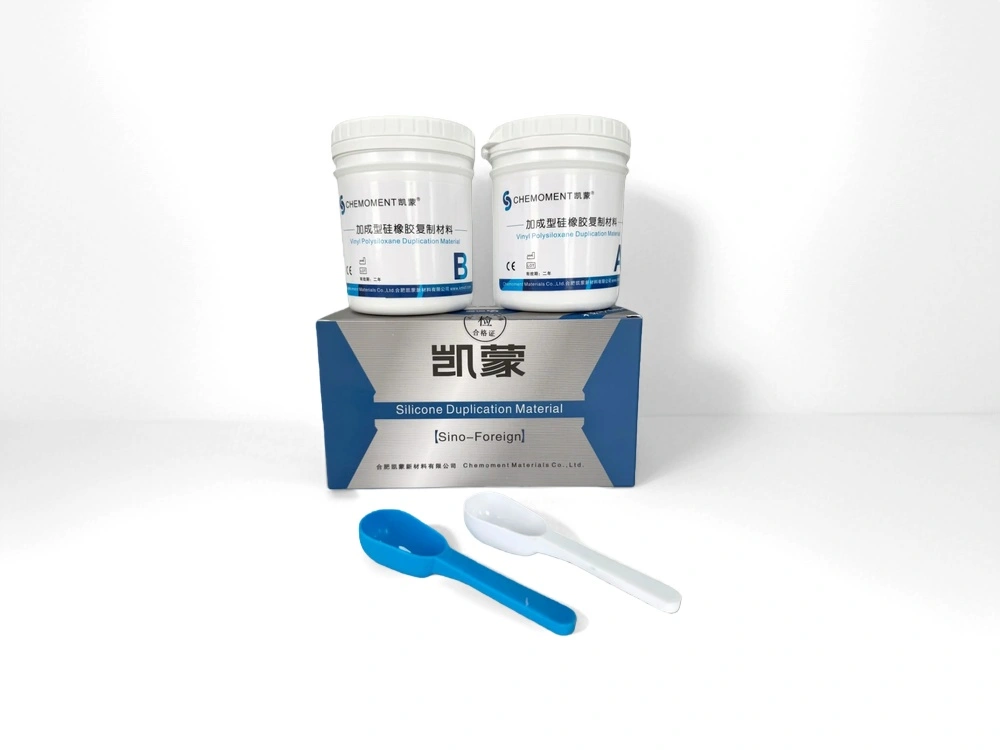
To overcome the dual challenges of tissue distortion and material shrinkage, the audiology team selected an advanced medical-grade addition-cure Silicone Ear Impression Material rather than standard industrial-grade putties.
The Chemoment formulation was chosen based on its specialized RTV-2 fluid dynamics. Its injection-grade, low-viscosity properties allow it to flow under minimal hydraulic pressure, capturing the exact, relaxed state of the epidermal lining without expanding the canal wall. Furthermore, its chemistry guarantees a linear shrinkage rate of less than 0.15%, ensuring that the physical dimensions captured chairside would remain identical during 3D laser scanning at the manufacturing laboratory weeks later.
4. Step-by-Step Clinical Implementation
The deep-canal impression procedure was executed via a cartridge-dispensed, dual-component automatic mixing system according to the following strict audiological protocol:
- Otoscopic Clearance & Blocking: A preliminary otoscopic examination confirmed a healthy tympanic membrane free of cerumen. A high-density foam otoblock (cotton-tipped tether) was gently positioned just past the second bend using a lighted impression pen to secure the tympanic boundary.
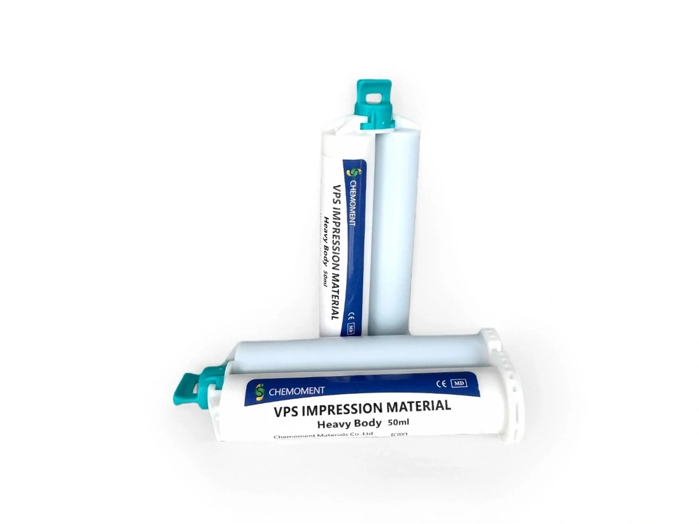
- Submerged Extrusion: The Chemoment 1:1 auto-mix cartridge was loaded into the dispensing gun. The mixing tip was placed directly against the otoblock, and injection began. The tip remained deeply submerged within the rising silicone matrix to prevent the introduction of air voids or seam lines.
- Passive Stabilization: The material was injected until it filled the concha and overfilled the helix rim. The patient was instructed to keep her jaw relaxed (neutral position) during the 4-minute setting phase to avoid dynamic jaw-joint movement errors.
- Tactile Cure Verification: After 4 minutes, a plastic probe confirmed that the flash material had transitioned into a smooth, elastic matrix with a calibrated final hardness of approximately 32 Shore A.
- Rotational Extraction: The impression was gently decoupled from the ear canal wall via anterior rotation and pulled out smoothly along the anatomical axis. Thanks to the material's high tear strength, the deep canal segment bypassed the undercuts completely intact without any marginal elongation.
5. Quantitative Outcomes & Long-Term Evaluation
The resulting impressions were shipped to a digital earmold facility for 3D scanning and shell replication. The integration of the advanced addition silicone system yielded significant quantifiable improvements over the patient's clinical baseline:
| Clinical Parameter | Previous Putty Baseline | Chemoment A-Silicone Outcome |
|---|---|---|
| Acoustic Feedback Threshold | Whistling triggered at >45 dB gain | Zero feedback at full prescription gain (+65 dB) |
| Daily Pain-Free Wear Time | Maximum 2 hours due to tissue pressure | 14+ hours (Continuous all-day wear) |
| Impression Tear / Remake Rate | 15% stretch deformation at canal bends | 0% (Flawless extraction across multiple trials) |
At the 6-month clinical review, the patient reported exceptional satisfaction, citing a total absence of acoustic whistling even during dynamic jaw movements like chewing or smiling. By switching to a low-pressure, dimensionally stable addition silicone supplier system, the clinic successfully eliminated lab remakes, optimized acoustical seal integrity, and provided a comfortable, long-term hearing solution.
For audiology networks, hearing aid manufacturers, or international medical distributors looking for premium ear impression materials complying with CE and FDA quality control standards, please reach out via our global B2B portal on the Contact Us page.